In the previous blog, I shared the story of my teammate’s experience with multi-directional shoulder instability, explained the types of shoulder instability and how they can present, and discussed who is likely to suffer from shoulder instability. In part two of this series, we will dive deeper into the anatomy of shoulder instability and explain where most treatments go wrong that prevent an ideal outcome!
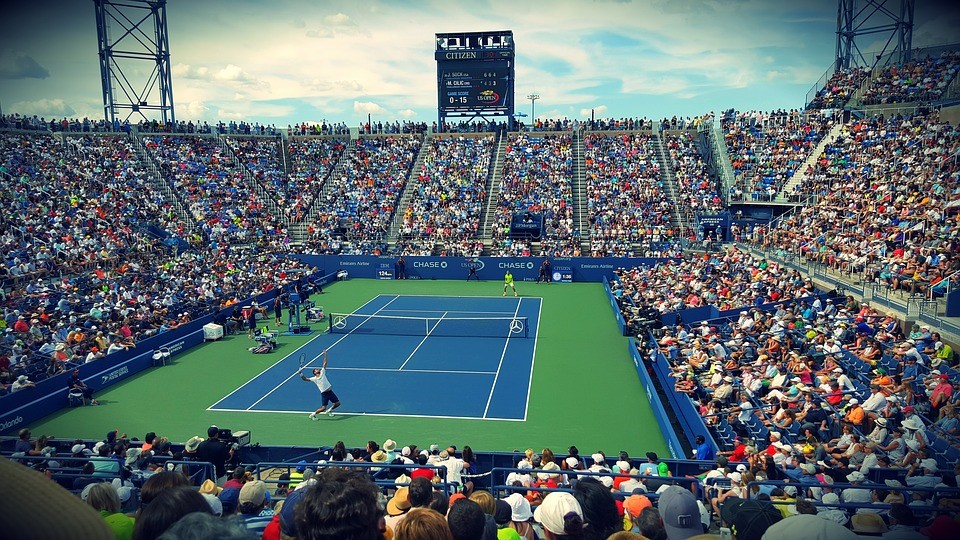
The glenohumeral or shoulder joint is an amazing joint for its’ ability to provide exceptional mobility, and yet still be strong enough to allow pitchers to throw 90+ MPH fastballs, tennis players to hit 130+ MPH serves, and gymnasts to vault multiple feet in the air. However, because of its’ ability to achieve a great deal of motion, the shoulder is the most dislocated joint in the body.
Attempting to reach such amazing feats can lead to a break down in the stability of the shoulder through overuse, insufficient muscle strength/musculature imbalance, or having poor bony anatomy that limits joint congruency and predisposes an athlete to instability.
Understanding the anatomy of the shoulder is imperative in properly diagnosing and treating the cause of shoulder instability. The shoulder has both passive and active stabilizers. Passive stabilizers include the bony structures, ligaments, labrum, and joint capsule, while the muscles surrounding the joint make up the active stabilizers.
The glenohumeral joint is considered a ball-and-socket joint consisting of the sphere shaped humeral head sitting in contact with the concave shaped glenoid fossa of the scapulae (shoulder blade). The humeral head has 3x the surface area as the tee-shaped glenoid fossa, which means there is relatively little contact area between the two bones. Without sufficient compression of humeral head into the fossa, there is a chance for humerus to roll out of the stability angle causing a dislocation of the joint.

The specific passive restraints that help keep the shoulder from dislocating include the superior, middle, and inferior glenohumeral ligaments, the coracohumeral ligament, the capsule surround the joint, a fibrocartilaginous labrum that surrounds and deepens the glenoid cavity, and negative pressure inside the joint creating a vacuum-like effect. Each of these passive structures can become compromised and lead to instability.
Ligaments and the capsule can become torn or stretched out through overuse. The labrum can become torn due to excessive translation/dislocation of the humeral head pushing up against it, or excessive pull from the long biceps tendon, which inserts into the labrum. Even the negative pressure within the joint can become disrupted from surgery. It is also possible to have the bony anatomy disrupted when the shoulder dislocates (Hill-Sachs or Reverse Hill-Sachs lesion).
Occasionally, the unstable shoulder will require surgery to repair torn ligaments, labral tears, tighten the capsule through plication, or fix bony/chondral lesions, but this is usually only done after an appropriate rehabilitation program has failed to control the patient’s symptoms.
The goal of a rehabilitation program is to improve the dynamic stability of the glenohumeral joint through proper training of the surrounding musculature. The rotator cuff is the primary dynamic stabilizer of the shoulder joint. It consists of four muscles (supraspinatus, infraspinatus, teres minor, and subscapularis) whose main function is to provide humeral head compression into the glenoid fossa through a synergistic relationship. The primary movers of the shoulder include the deltoid, pectoral major, latissimus doris, and teres major.
The supraspinatus assists with superior capsular stability, the subscapularis provides anterior stability, while teres minor and infraspinatus provide posterior capsular stability. The long head of the biceps tendon can assist with anterior shoulder stability as it runs across the front of the humeral head.
Since the glenoid fossa is part of the scapula, it is also important to ensure proper stabilization and movement of the shoulder blade. The scapulae are only attached to the rest of the skeleton via the clavicle and sternoclavicular joint. It is mostly held in place by 17 muscles, and if these muscles do not provide good dynamic stability, it is even more difficult to provide stability at the glenohumeral joint.
A good rehabilitation program will also look at proper strength, muscular balance, and scapulothoracic rhythm to ensure there is a stable base for the shoulder to move upon. This is one area that rehabilitation treatments may miss leading to suboptimal results.

Rehabilitation programs that are successful at treating shoulder instability will first assess seven key factors: onset of pathology, degree of instability, frequency of dislocation, direction of instability, associated pathologies, neuromuscular control, and previous activity level. This will help guide proper treatment for which muscles to strengthen in order to help restore stability to the shoulder. A comprehensive program will include restoring shoulder ROM if needed, balance capsular mobility, improve neuromuscular control and proprioception, and provide muscular strength and endurance to appropriate rotator cuff and scapular muscles.
Unfortunately, many physical therapists do not have time to fully assess all of these factors during their evaluation. This often leads to general shoulder programs that aren’t specific to the type of shoulder instability and often provide outcomes not sufficient for a full return to sport.

This is EXACTLY why we do things differently at Competitive Edge Physical Therapy. We provide the longest treatment times in the Bay Area so that we can go through a comprehensive shoulder examination and ensure a proper diagnosis and treatment plan that will lead to the most successful outcome!!
If you would like to learn more about how we differ from other PT clinics, how we utilize cutting edge technology for improving movement patterns and recovery time, or just have questions regarding shoulder instability, feel free to give our office a call, send us an email, or stop by our office located in San Jose!
Citations:
- Lynch, B., Ridge-Hankins, T., Vyas, D. “Shoulder Instability: A Review of Anatomical and Biomechanical Considerations, Prevalence, and Diagnosis as well as Nonoperative and Operative Management”. Alternative Special Topics: Innovations in Practice. Independent study Course 25.3.1.
